Case 8: Topcon 3D OCT OS
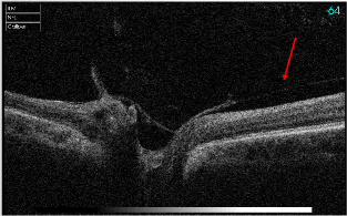
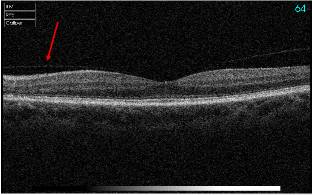
Notice structure attached to retina extending to optic nerve and periphery.


• 72-year-old presented with vague symptoms in left eye but with 20/20 VA and a nonspecific reduction in sensitivity on visual field.
• Patient’s optic nerve in left eye was judged to have blurred disc borders.
• MRI was normal, complete blood work was similarly normal and ruled out temporal arteritis.
• Subsequent to MRI, an OCT of the disc was performed and revealed a dramatic vitreal papillary traction which was likely responsible for the visual field defect and blurred disc borders.
• Over several months all the findings slowly resolved without treatment.


