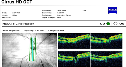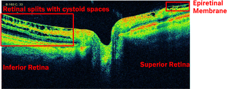

The 5 line vertical raster scans (high resolution) reveal cystoid spaces and several retinal splits (schisis) inferior to the optic nerve head and an epiretinal membrane superior to the optic nerve head. The magnified scan below corresponds to the blue vertical line in the fundus image to the left. The four other scans represent the green vertical lines as indicated.


