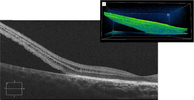
Case 2: High Definition OCT and Topcon 3D Image of Right Eye
Case 2: The horizontal OCT section and 3D image document the detachment of the neurosensory retina temporal to the macula. Note the cystic type gaps on either side of the outer plexiform layer.


