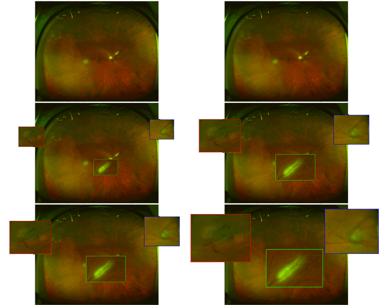
Case 1: Optomap® Color Fundus Image OD

52-year-old white female complained of reccnt onsct ot a largc floatcr in thc rignt cyc. Thc optomap® image revealed a superior temporal horseshoe tear with a small hemorrhage (red square) and a superior nasal retinal hole or small tear (blue square). BIO confirmed these two findings. She was also a glaucoma suspect based on large cups and subtle visual field loss but her pressures were always measured in the mid teens. Note the rectangular white lesion (green square) perhaps represents a small area of medullation. BCVA was 20/25 OD and 20/25 OS.


