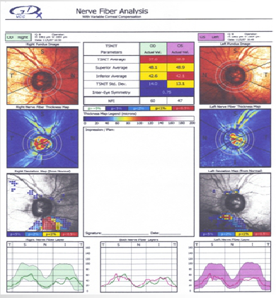
Retinal nerve fiber layer abnormalities OU as shown with GDx. The Deviation Map reveals an inferior loss OU which has progressed somewhat OD. Both the fields and the GDx shown were obtained prior to the recent vein occlusion.
Learn more about GDx and OCT RNFL interpretation.*
*The Retinal Nerve Fiber Layer in Clinical Practice is available at: http://www. Iulu.com


