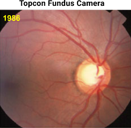
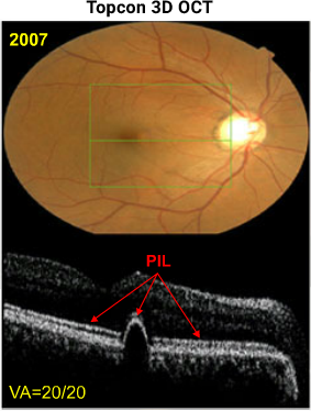
Left image: Photo of right fundus back in 1986 when patient presented with CRVO in the fellow eye.
Right images: On routine follow-up in 2007, asymptomatic patient with 20/20 VA presented with a pigment epithelial detachment (PED) slightly nasal to the fovea OD which is documented with OCT. The C/D ratio appears larger in 2007 than in 1986 likely due to glaucoma progression over the two decade period.


