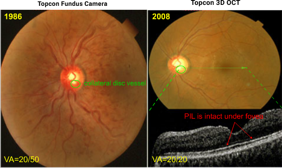
The fundus photo OS demonstrated the presence of a collateral disc vessel in 1986. The hemorrhages absorbed over the next year or so.
Two decades later, the SD OCT demonstrates an intact PIL under the fovea. A normal PIL, such as in this eye at this time, is highly correlated with normal VA.1 In addition, the collateral disc vessel has regressed.


