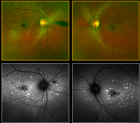
Case 5: Optomap® pIus with ResmaxTM Magnification OD and OS
KEY:
Color Optomap
Auto Fluorescence
In these Resmax images, the dots, spots and íish tail shaped lesions demonstrate marked hyper AF in each eye. The right eye exhibits a PVD nasal to the disc but in different positions in the two OD images which proves that the lesion is not within the retina.


