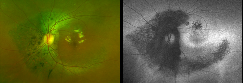
Case 6: Optomap® Color Fundus and AF Comparison OS

In the color fundus Image, note the hard exudates (In the macula) whIch are typIcally In the outer plexIform layer. Hard exudates In Henle’s FIber Layer tend to form a macular star. As expected, hard exudates are virtually Invisible with AF, sInce AF Is essentially a RPE phenomena.


