

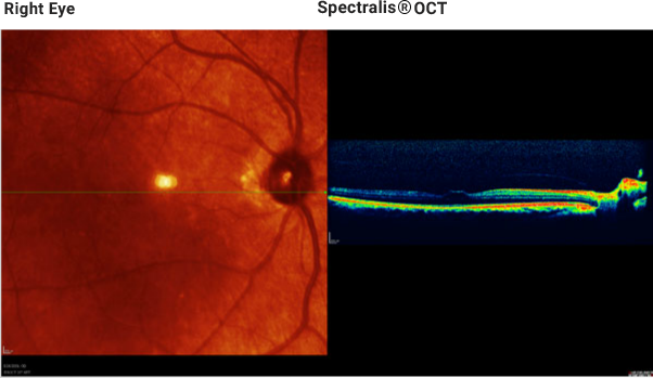
The horizontal scan through the macula and optic nerve head demonstrates the attachment of the posterior vitreous at the macula and the optic nerve head.
Note: The presence or absence of the PIL is generally better documented with gray scale imaging.