

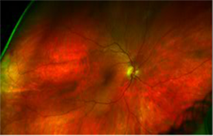
Figure A – Composite view of the right eye.
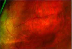
Figure B – Magnified view of right eye.
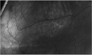
Figure C – Magnified view of right eye using the green separation.
Asymptomatic 36 year old female presented with a far peripheral anastomosis in the right eye temporally (as well as a small hemorrage above the AV abnormality). Such findings may be congenital, but can be acquired due to diseases such as sickle cell anemia, sarcoid and leukemia. In this case, no etiology of AV anastomosis has been identified.
Panoramic Ophthalmoscopy: Optomap Images and Interpretation is available at:
http://www.slackbooks.com/view.asp?SlackCode=67808