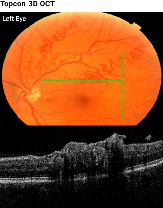
The green box is 6 mm x 6 mm and contains 128 horizontal sections.
The retina is elevated around the AV malformation. “Shadowing” of deeper structures prevent detailed assessment of the PIL and other layers.
When both the PIL and RPE are not present, no conclusion can be reached about the integrity of the PIL in that area shadowed by the abnormal vessels.


