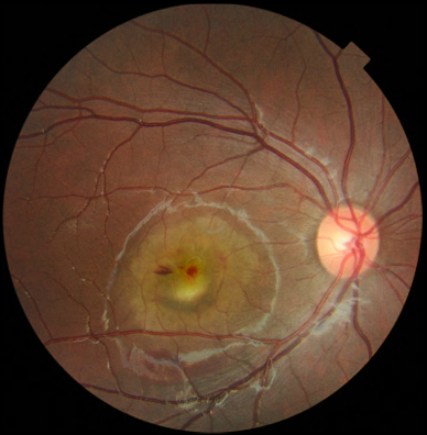
Case 1: Topcon 3D OCT Color Fundus Image OD

Case 1: A 6-year-old female was referred for her first eye exam after failing a school screening one week earlier. Her BCVA measured 20/200 OD and 20/100- OS. IOPs were 16 mmHg OU. Fundus examination revealed bilateral chorloretinal lesions with intraretinal hemorrhaging. The yellow white “sunny side up egg” lesion is beginning to scramble in the right eye.


