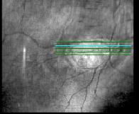
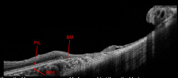
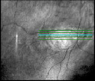
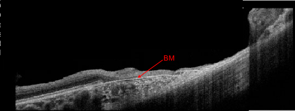
Case 1: Cirrus™ HD-OCT Horizontal 5-Line Raster Scan Images OS


All the retinal layers appear normal before reaching the retinal lesion. As the retinal layers reach the border of the lesion several of them appear to “collapse”. An OCT section through the lesion reveals several absent retinal layers down to Bruch’s membrane (BM). BM is typically not visualized in OCT images unless the overlying retinal layers, such as the RPE, are missing.


