OCT and Visual Field Limitation: Vitreous Floaters
Vitreous floaters and posterior vitreous detachments are more common in myopic patients. Posterior vitreous detachments complicate OCT and visual field measurements because when the floaters move it obstructs the imaging. One way to avoid this limitation is to have the patient look up to move the floater out of the way for a moment to capture a more accurate photograph.

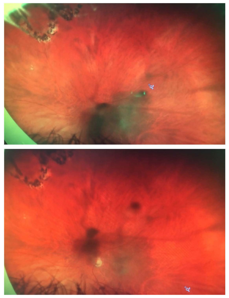
Optos color fundus photographs that demonstrate a very large floater in a -6.00 D myope
that moves and complicates visual field and OCT images.


