Orbital Disorders
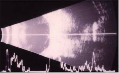
Since the orbit, unlike the retina, cannot be viewed directly, ultrasonography is invaluable in the detection of abnormalities that may be overlooked by ophthalmoscopy. In orbital diagnosis, the B-mode is ideal for the identification and delineation of orbital anomalies. A-mode analysis and dynamic scanning add information to the pattern obtained from the B-mode.
Recall that in the globe, abnormalities appear white or gray against a black background. Orbital abnormalities, on the other hand, are displayed as dark defects against a white background (the acoustically dense orbital fat). Abnormalities in the orbit are divided into three categories: (1) mass lesion, (2) foreign body, (3) inflammatory change.

B-scan of the orbit demonstrating a mass in the orbit which is close proximity to the optic nerve. Of interest, this patient had an orbital tumor removed several months earlier and the apparent “regrowth” of the tumor turned out to be only scar tissue which remained unchanged over a several year follow-up.


