Orbital Disorders

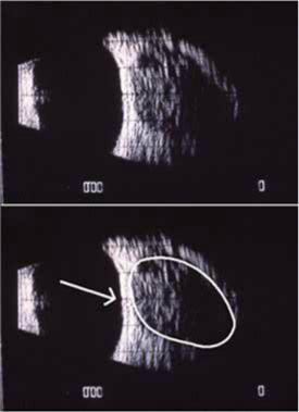
The patient whose ultrasonogram is pictured to the left complained of reduced vision in her right eye for 3 to 4 years, with lid swelling for the last 2 to 3 years. Pupillary testing revealed a Marcus Gunn pupil in that eye. Motilities of the right eye were restricted in superior gaze. Visual acuity was 20/200, and the patient had proptosis of the right eye. Retinal examination revealed pigmentary changes in the posterior pole.
The B-mode ultrasonogram shows a large retro-bulbar mass. It was oval, with little or no evidence of the posterior wall seen in the scan. Histopathologic examination of the mass after its removal confirmed it to be a meningothelial meningioma of the optic nerve.


