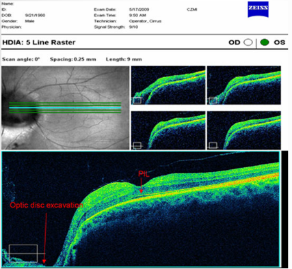
The 5 line horizontal raster scans (high resolution) reveal the appearance of the optic disc excavation and macular region. The magnified scan below corresponds to the blue horizontal line in the fundus image to the left. The four other scans represent the green horizontal lines as indicated. The PIL is intact under the foveal pit and predicts normal VA if the retinal nerve fiber layer in the papillo-macular bundle is not compromised.


