It Is well recognized that optomap® FA reveals select abnormalities that are not visible with Ophthalmoscopy or fundus photography. Optos optomap® FA has the advantage of viewing virtually the entire fundus at once and reveals findings typically missed with standard FA. Three Optos products exist at present: P200, the P200C and the P200MA. Retinal specialists typically prefer the P200MA which has the same increased resolution as the P20 C but also incorporates optomap® FA capabilities.
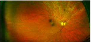
This is an example of color optomap® imaging and optomap® FA in a different patient. Note the peripheral retinal abnormality in the temporal retina of the right eye in the color image and the optomap® FA. In addition, the optomap® FA of the left temporal retina is also not normal. This “mysterious” case will be presented in detail in an upcoming Retina Revealed – stay tuned!


