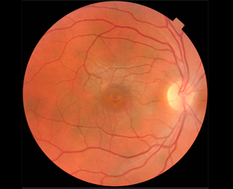
Case 1: Signature case of MSI detecting large choroidal lesions
invisible to ophthalmoscopy, OCT, and MRI

A 25 year old female presented with BCVA 20/20 OD and OS. She was concerned about her eyes because she was told by her neurologist that patients with her disorder, neurofibromatosis type 1, sometimes develop visual problems. Previous eye exams failed to reveal any unusual findings. The fundus through a dilated pupil was considered to be unremarkable with direct ophthalmoscopy and with indirect ophthalmoscopy (with a 78D lens and with a BIO). Fundus photography was also judged to be unremarkable in both the right and left eye.


