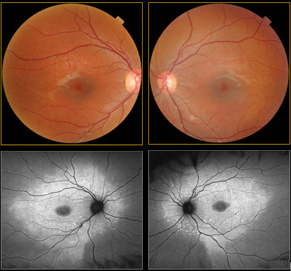
Case 1 (8 yo son): Fundus Photography
KEY:
Color Optomap
Red Separation
Green Separation
Auto Fluorescence
A comparison of standard fundus photos to AF images clearly reveals unmistakable abnormalities in the AF images only. Note that the small hyper AF lesions are often not round and many are fish tail or pisciform in shape, suggesting perhaps the fish tail lesions in Stargardt’s Disease with fundus flavimaculatus. (#RR14) The symmetry of the AF findings are quite typical of inherited retinal degenerations. (#RR54 part1 & #RR54B part2)


