Vitreomacular Traction Syndrome – page 16 of 26
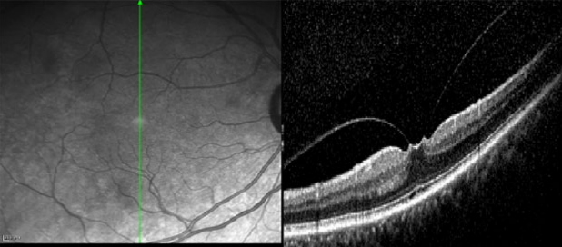
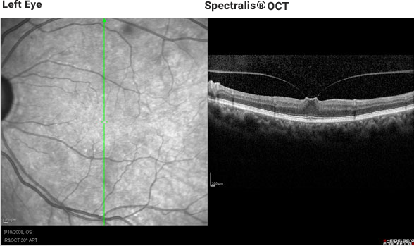
The vertical section through the fovea depicts vitreal macular traction, which distorts the presence of normal foveal pit (see page 21).
Sponsored by:


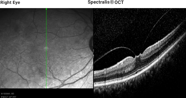
The vertical section through the fovea depicts vitreal macular traction, which distorts the presence of normal foveal pit (see page 21).
The vertical section through the fovea depicts vitreal macular traction, which distorts the presence of normal foveal pit (see page 21).
Heidelberg Spectralis OCT 2 dimensional movie which is composed of 19 horizontal sections beginning from the bottom and going up. Note: The movie can easily be paused or replayed by moving the mouse over the bottom of the black box to reveal a control panel. Left Eye Heidelberg Spectralis® OCT 2 Dimensional Movie
Example 3: VMT Syndrome in another patient History/Chief Complaint 76 year old Hispanic female with glaucoma and a previous history of filtering surgery OU, diabetes without retinopathy and systemic hypertension. Clinical Findings BCVA: 20/20 OD and 20/25 OS Filtering blebs OU and clear and centered PC IOLs OU. Fundus Exam: Large cups OU and
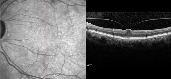
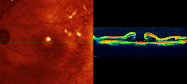
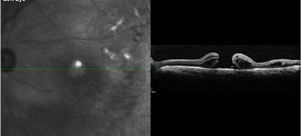
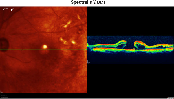
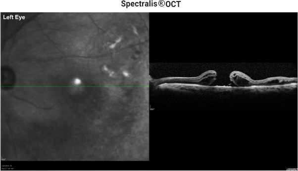
The horizontal section through the fovea depicts vitreal macular traction, which distorts the presence of a normal foveal pit (see page 21).
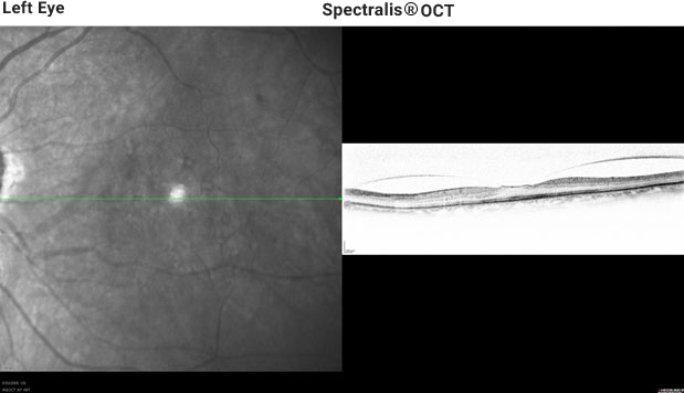
The horizontal section through the fovea depicts vitreal macular traction, which distorts the presence of normal foveal pit (see page 21).
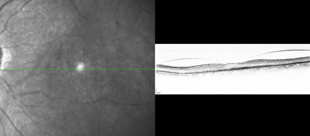
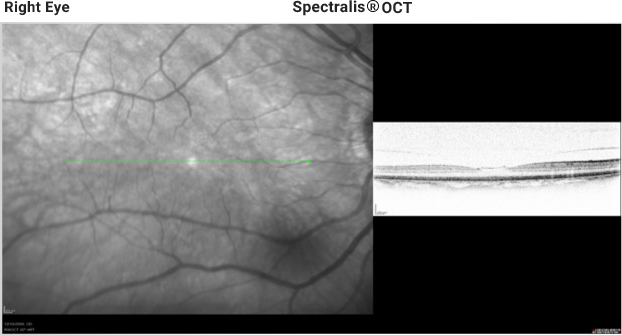
Example 2: VMT Syndrome History/Chief Complaint 74 year old Hispanic female with early glaucoma under treatment. Clinical Findings BCVA: 20/40 OD and 20/40 OS Moderate nuclear cataracts OU Fundus Exam: Unremarkable VA reduction commensurate with cataract OU. Spectralis™ HRA+OCT Image OCT image: VMT syndrome with intact PIL (see page 21) in each eye.* *The
The OCT and fundus image below is the section demonstrated on the previous page, but depicted in color.
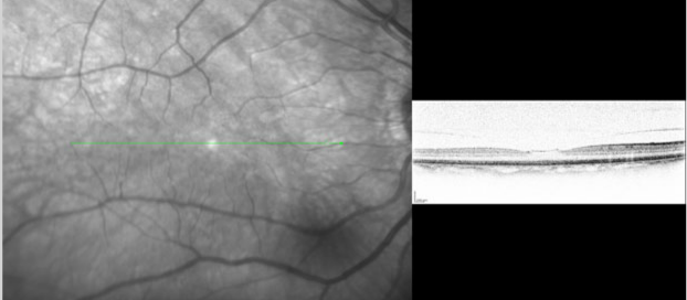
Spectralis® OCT depicting a horizontal section OS through the fovea reveals a full thickness macula hole without evidence of VMT (Grade 4). The PIL (see page 21) is not observable at the base of the hole. Cystoid spaces in a thickened retina surround the hole.
Fluorescein angiogram was obtained after the patient developed vein occlusions. The FA below demonstrates the presence of leakage secondary to an inferior temporal branch vein occlusion OS. Left Eye Heidelberg Spectralis OCT 2 dimensional movie which is composed of 19 horizontal sections beginning from the bottom and going up. Note: The movie