

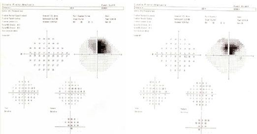
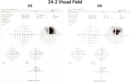
Stargardt Plus Glaucoma – Page 30 of 30
Suggested Readings E. Ergun, B. Hermann, M. Wirtitsch, A. Unterhuber, TH Ko, H. Sattmann, C. Scholda, JG Fujimoto, M. Stur, W. Drexler 2005 Assessment of Central Visual Function in Stargardt's Disease/ Fundus Flavimaculatus with Ultra-high Resolution Optical Coherence Tomography. Investigative Ophthalmology & Visual Science. 46(1): 310-316 GA Fishman, EM Stone, S. Grover, DJ Derlacki,