

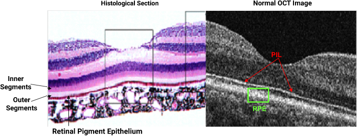
Suspicious Nevus – page 23 of 23
References: Augsburger J.J., Correa Z.M., Trichopoulos N., Shaikh A. Size Overlap between benign melanocytic choroidal nevi and choroidal malignant melanomas. Investigative Ophthalmology and Visual Science 2008; 49 (7): 2823-2827 Abramson, D. (Personal communication, May 2009) Shields C.L. The hunt for the secrets of uveal melanoma. Clinical and Experimental Ophthalmology 2008; 36:277-280 Shields C.L., Shields J.A.,