AV Malformation (AVM) – Page 19 of 22
The following is a list of links to the companies and contributors of Congenital Arteriovenous Malformation :
Sponsored by:


The following is a list of links to the companies and contributors of Congenital Arteriovenous Malformation :
Comments and Conclusions Con’t Arteriovenous communications of the retina can be classified into three groups4: - Group 1-The retina shows interposition of an abnormal capillary plexus between the major vessels, mostly without visual symptoms. - Group 2-The retina reveals direct arteriovenous communications without interposition of capillaries. Patients have few visual symptoms, In addition, associated cerebral
Comments and Conclusions Arteriovenous malformations result from persistence of the embryonic vascular pattern that exists before the arteries, capillaries and veins differentiate during the second month of gestation.4 It is a rare, unilateral nonhereditary disorder with variable visual impairment which depends on the severity of the anomaly. 4 A complete literature search of retinal arteriovenous
Ocular complication of AV Malformation Although usually non-progressive, retinal arteriovenous anomalies can be associated with the following complications:3 Intraretinal macular hemorrhage Intraretinal hemorrhages outside the macula Neovascular glaucoma Vitreous hemorrhage Central and branch retinal vein occlusion Retinal exudation
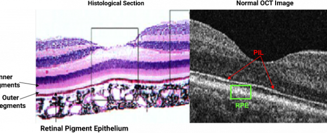
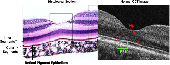
Histological Section as Compared to the OCT Image Although the photoreceptor integrity line, or the PIL (defined as the junction between the inner and outer segments) is barely visible in most histological sections, it is highly prominent in normal SD OCTs. The PIL, as shown above should be continuous throughout the entire scan in
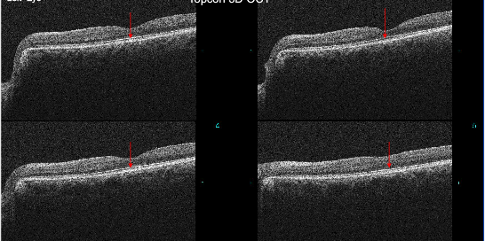
The four consecutive OCT scans above reveal the presence of a normal and intact PIL under the foveal pit OS and as well as under the entire macula. The images obtained correlate with BCVA acuity of 20/15. Although both eyes have an epiretinal membrane in the macula, the ERM in the OD is slightly larger
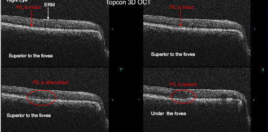
Above are four consecutive horizontal sections through the macula OD. The PIL is near normal and relatively intact superior to the fovea and is absent under the foveal pit. By looking above the letters to be read on the Snellen Chart, the patient is placing the letters to be read on retinal points superior to
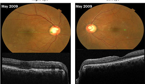
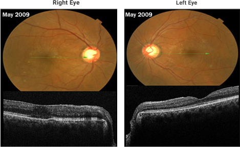
Follow-up of the vein occlusion patient from previous page: The BCVA was 20/30 OD and 20/15 OS. Ophthalmoscopy revealed large cups due to glaucoma OU and an epiretinal membrane 0D>0S. The vein occlusions OD appear to be resolving quite well but the patient is still symptomatic in the right eye. He complains about persistent
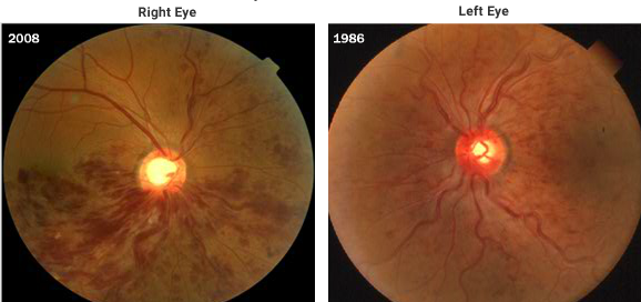
Image Gallery - Vein Occlusions The images above are from a patient who had a CRV occlusion OS more than two decades ago and a recent inferior hemi-central retinal vein occlusion OD. An additional superior nasal vein occlusion months earlier gives the appearance of a 3 quadrant vein occlusion OD.(See Case 5- Vein Occlusion)
Figure A - Composite view of the right eye. Figure B - Magnified view of right eye. Figure C - Magnified view of right eye using the green separation. Asymptomatic 36 year old female presented with a far peripheral anastomosis in the right eye temporally (as well as a small hemorrage above the AV abnormality).