Case #5 – Vein Occlusion

Three Quadrant Retinal Vein Occlusion – Page 9 of 19

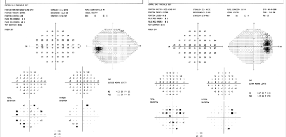
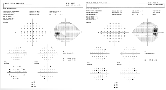
Visual field loss is most likely due to the POAG but has not progressed for over a decade. Goldmann lOPs have been below 20 consistently with good compliance to medications. The C/Ds are large OU but have not changed over the last decade (but have increased over the two decade period). The full medical workup

Three Quadrant Retinal Vein Occlusion – Page 9 of 192021-11-22T23:07:14+00:00

Three Quadrant Retinal Vein Occlusion – Page 8 of 19

The fundus photo OS demonstrated the presence of a collateral disc vessel in 1986. The hemorrhages absorbed over the next year or so. Two decades later, the SD OCT demonstrates an intact PIL under the fovea. A normal PIL, such as in this eye at this time, is highly correlated with normal VA.1 In

Three Quadrant Retinal Vein Occlusion – Page 8 of 192021-11-22T23:07:04+00:00

Three Quadrant Retinal Vein Occlusion – Page 6 of 19

Right images: Symptomatic patient presents with blurred vision OD in 2008 with a new inferior hemi-central retinal vein occlusion (or hemiretinal vein occlusion) in addition to the previous, partially absorbed superior nasal vein occlusion. Full (repeated) systemic workup was noncontributory (see page 16). Selective laser trabeculoplasty (SLT) successfully lowered the lOPs to the lower

Three Quadrant Retinal Vein Occlusion – Page 6 of 192021-11-22T23:06:40+00:00

Three Quadrant Retinal Vein Occlusion – Page 5 of 19

Left image: Photo of right fundus back in 1986 when patient presented with CRVO in the fellow eye. Right images: On routine follow-up in 2007, asymptomatic patient with 20/20 VA presented with a pigment epithelial detachment (PED) slightly nasal to the fovea OD which is documented with OCT. The C/D

Three Quadrant Retinal Vein Occlusion – Page 5 of 192021-11-22T23:06:31+00:00

Three Quadrant Retinal Vein Occlusion – Page 4 of 19

Panoramic Ophthalmoscopy as provided by optomap® is the Ideal imaging approach to document hemorrhages in three quadrants extending from the fovea to beyond the equator. Note the absence of hemorrhages in the entire superior temporal quadrant. Serial images over time with optomap® are uniquely suitable to document either progression or regression of the hemorrhages at

Three Quadrant Retinal Vein Occlusion – Page 4 of 192021-11-22T23:06:22+00:00

Three Quadrant Retinal Vein Occlusion – Page 2 of 19

History/Chief Complaint 52 year old, moderately myopic Hispanic male presented with blurred vision in the right eye for one week. Several months earlier, a superior nasal (SN) branch vein occlusion OD was observed. About 22 years ago, a CRVO was diagnosed in the OS. The full work-up at the time was negative and VA

Three Quadrant Retinal Vein Occlusion – Page 2 of 192021-11-22T23:06:05+00:00

Three Quadrant Retinal Vein Occlusion – Page 1 of 19

Although a branch retinal vein occlusion is common in the elderly population with underlying disorders such as systemic hypertension, multiple and recurrent vein occlusions in young healthy adults are rare. This week's case concerns a young healthy adult whose first documented vein occlusion (CRVO OS) occurred at age 30. More than two decades later,

Three Quadrant Retinal Vein Occlusion – Page 1 of 192021-11-22T23:05:56+00:00