“Morning Glory” Disc Coloboma – Page 17 of 27
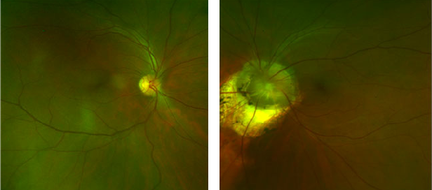
Figure A: Coloboma inferior to disc. Figure B: Coloboma of the disc and also of the retina and choroid inferior to the disc.
Sponsored by:


Figure A: Coloboma inferior to disc. Figure B: Coloboma of the disc and also of the retina and choroid inferior to the disc.
Optos ResMax™ images above demonstrate the dramatic difference in the size of the optic nerve heads. Simultaneous viewing of both eyes is easily performed with the optomap® software. Note: The images above were obtained at exactly the same magnification
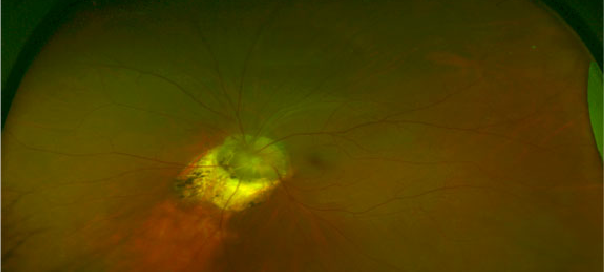
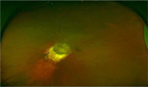
The optomap® color fundus image below represents a panoramic view of approximately 200 degrees x 150 degrees of the fundus and reveals the optic disc anomaly without any other abnormalities. Panoramic Ophthalmoscopy: Optomap® Images and Interpretation
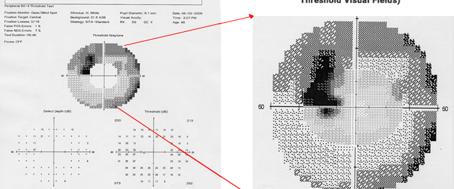
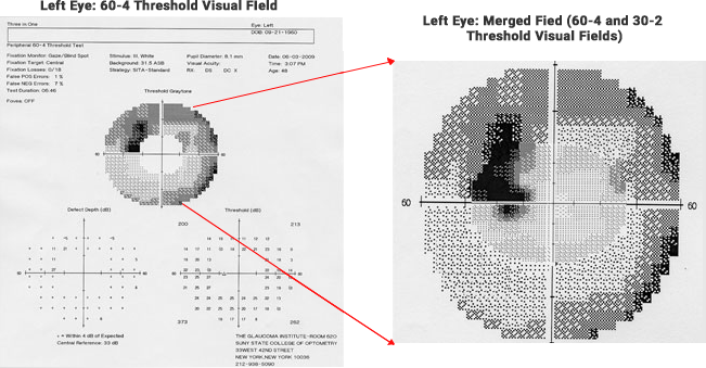
Zeiss Humphrey Visual Fields The 60-4 VF tests an annulus of points from 30 degrees to 60 degrees from fixation and no points within the 30 degrees. The image on the right represents the merged results of the two VFs into one image. (This merge of the central 30 degree field with the peripheral field
Zeiss Humphrey 30-2 Threshold Visual Fields
Figure A: ILM-RPE Figure B: Cirrus segmentation of the internal limiting membrane (ILM) Figure C: Segmentation of the retinal pigment epithelium (RPE) Horizontal folds are present in the ILM-RPE (Figure A) image and also in the ILM segmentation image (Figure B) but not in the RPE segmentation image
The 5 line horizontal raster scans (high resolution) reveal the appearance of the optic disc excavation and macular region. The magnified scan below corresponds to the blue horizontal line in the fundus image to the left. The four other scans represent the green horizontal lines as indicated. The PIL is intact under the
Zeiss Cirrus HD OCT 2 dimensional movie which is composed of 200 horizontal sections (Macular Cube 200 x 200) beginning from the top and going down. All sections are contained within the yellow box as demonstrated. Note: The movie on can easily be paused or replayed by use of the control panel on the
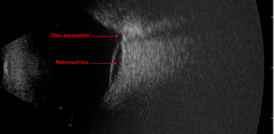
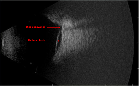
The DGH B-scan Scanmate above is a vertical slice through the optic nerve head and the retinoschisis inferior to the optic nerve head.* 8-Scan for Beginners is available at: http://www.lulu.com